69) Ford, T. L., 2009, How to Draw Dinosaurs: The dinosaurs with a bad name...Oviraptorids, Part 1: Prehistoric Times, no. 90, p. 18-19.
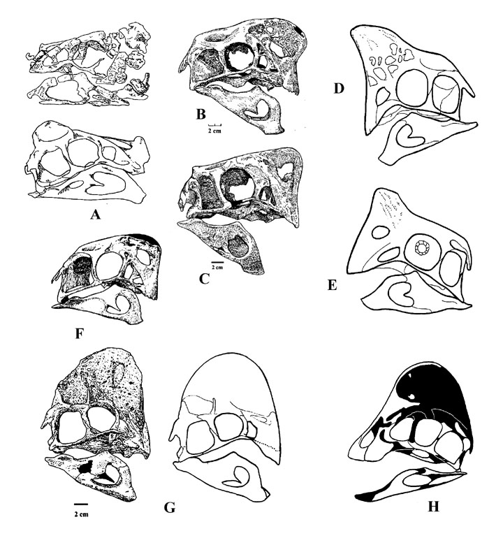
Figure 1) Crested Oviraptorosauria; A) Oviraptor philoceratops AMNH 6517; B) Oviraptor nova GI SPS No. 100/42; C) Nemegtomaia barsboldi GIN 100121 12; D) Oviraptor nova (Gaston); E) Oviraptor nova (Gaston); F) Citipati osmolskae IGM 100/978; G) Rinchenia mongoliensis GI SPS No. 100/32 (after Barsbold, and reconstructed skull); H) Chirostenotes sp (Triebold specimen, after Scott Hartman)
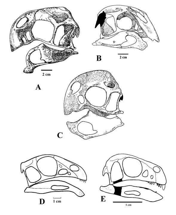
Figure 2) Crestless Oviraptorosauria; A) Conchoraptor gracilis GKH
PST No. 100/20; B) Khaan mckennae IGM 100/1127; C) Ingenia yanshini GI SPS
No. 100/30;
D) Caudipteryx zoui NGMC 97-4-A & NGMC 97-9-A; E) Protarchaeopteryx robusta
= Incisivosaurus gauthieri IVPP V13326.
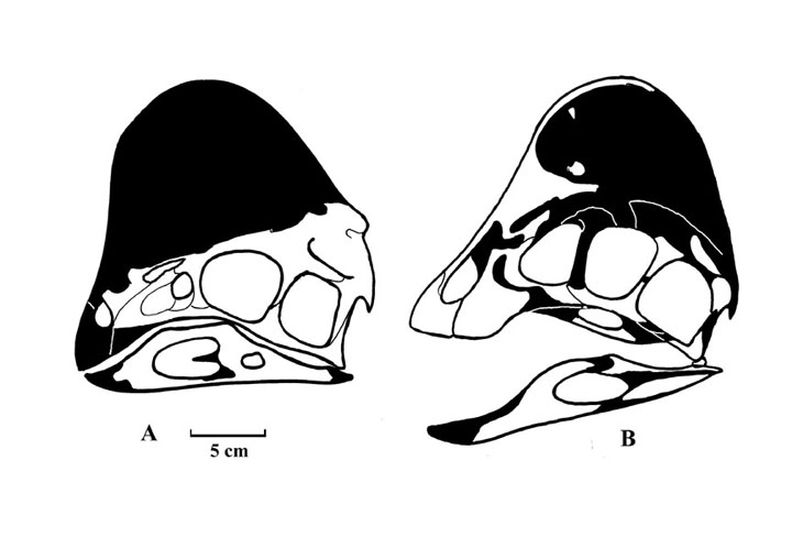
Figure 3) New interpretation of Oviraptor using Chirostenotes as a template; A) Oviraptor; B) Chirostenotes.
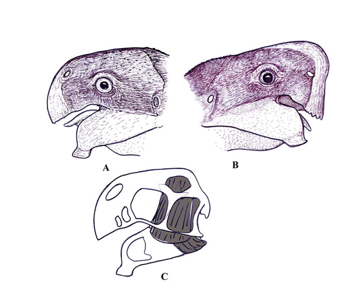
Figure 4) Head reconstructions of Oviraptorosauria; A) Conchoraptor
gracilis;
B) Citipati osmolskae; C) Jaw muscles of Conchoraptor gracilis.
70) Ford, T. L., 2009, How to Draw Dinosaurs: The dinosaurs with a bad name...Oviraptorids, Part 2: Prehistoric Times, no. 91, p. 18-19.
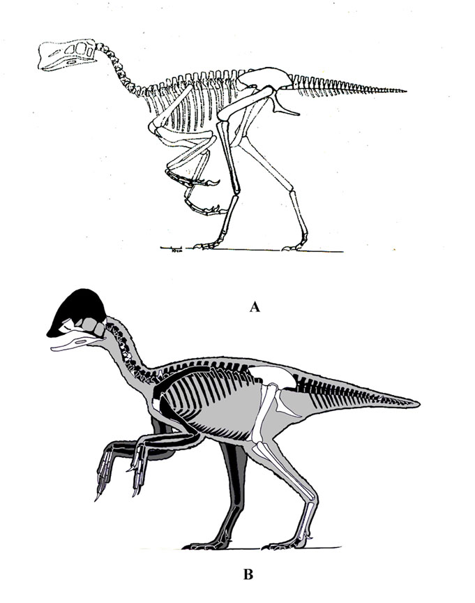
Figure 1). A) my first reconstruction of Chirostenotes; B)
my new interpretation.
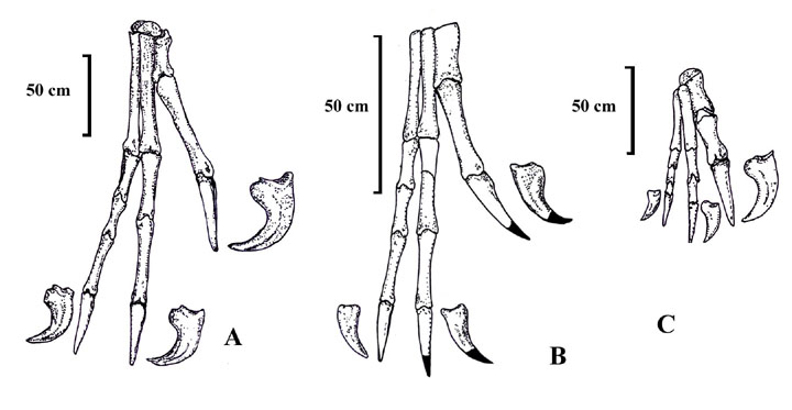
Figure 2). Hands of Oviraptorosauromorphs; A) Oviraptor philoceratops; B) Conchoraptor gracilis; C) Ingenia yanshini.
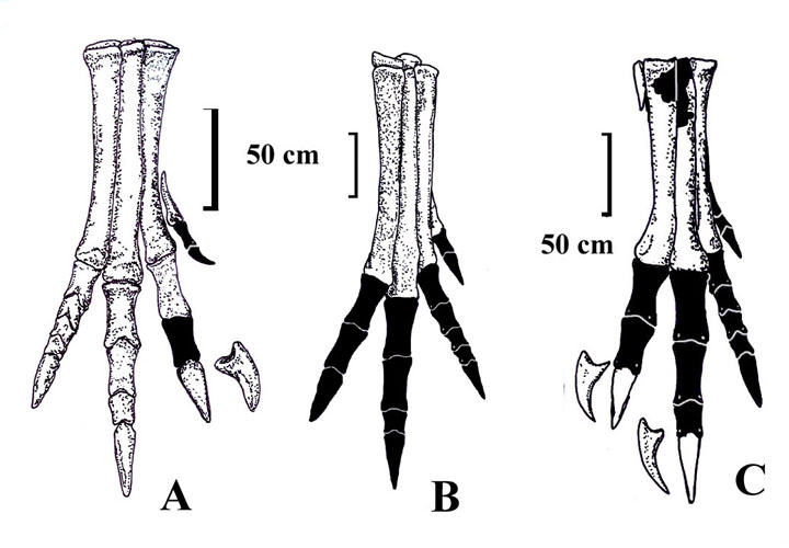
Figure 3) Feet of Oviraptorosauromorphs; A) Ingenia yanshini; B) Oviraptor philoceratops; C) Conchoraptor gracilis.
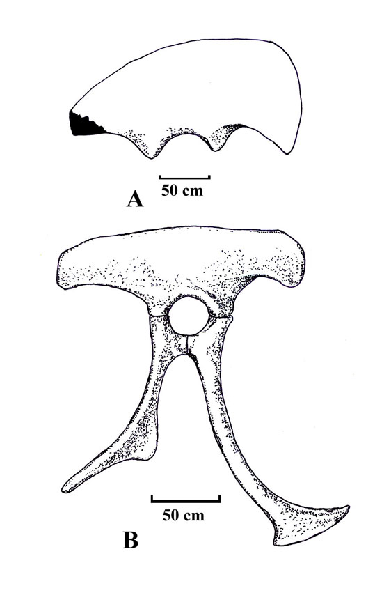
Figure 4). Pelvis and ilia of oviraptorosaurs. A) Rinchenia mongolensis; B) Ingenia yanshini.
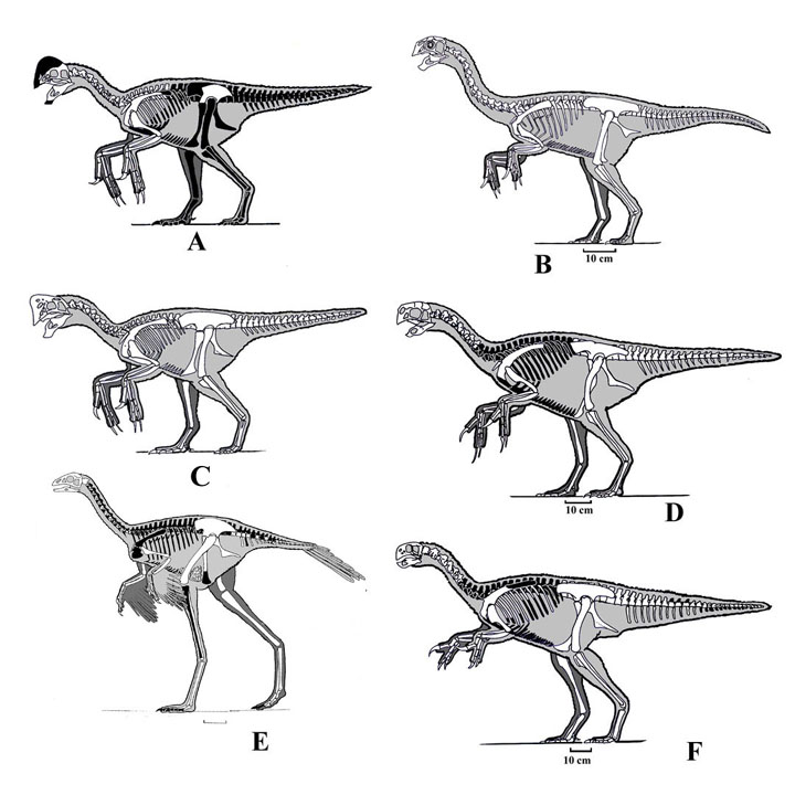
Figure 5) Oviraptorosauromorph skeletons: A) Oviraptor philoceratops; B) Khaan
mckennae; C) Citipati osmolskae; D) Conchoraptor gracilis; E) Caudipteryx
zoui;
F) Ingenia yanshini.
71) Ford, T. L., 2009, How to Draw Dinosaurs: Bulking up Sauropods (and dinosaurs in general): Prehistoric Times, n. 92, p. 18-19.
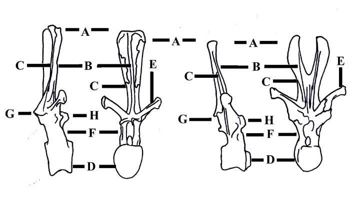
Figure 1) Diagram of vertebra of Dicraeosaurus; A) rugosity on the top of the neural spine; B) neural spine; C) spdl (spinodiapophyseal lamina); D) centra; E) diaphyoses; F) neural arch; G) postzygophyseis; H) prezygophyseis.
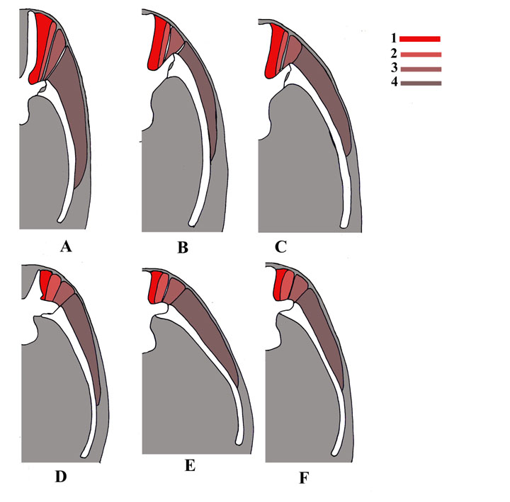
Figure 2) Cross section of the body of sauropods showing the muscles; A-C Dicraeosaurus, D Diplodocus, E-F Apatosaurus; A) Dicraeosaurus anterior dorsal vertebra; E) Dicraeosaurus middle dorsal; F) Dicraeosaurus posterior dorsal; D) Diplodocus anterior dorsal vertebra; E) Apatosaurus mid dorsal; F) Apatosaurus posterior dorsal; color identification of muscles; 1) m transsp med; 2) m. transsp lat; 3) m long d; 4) m ilcost.
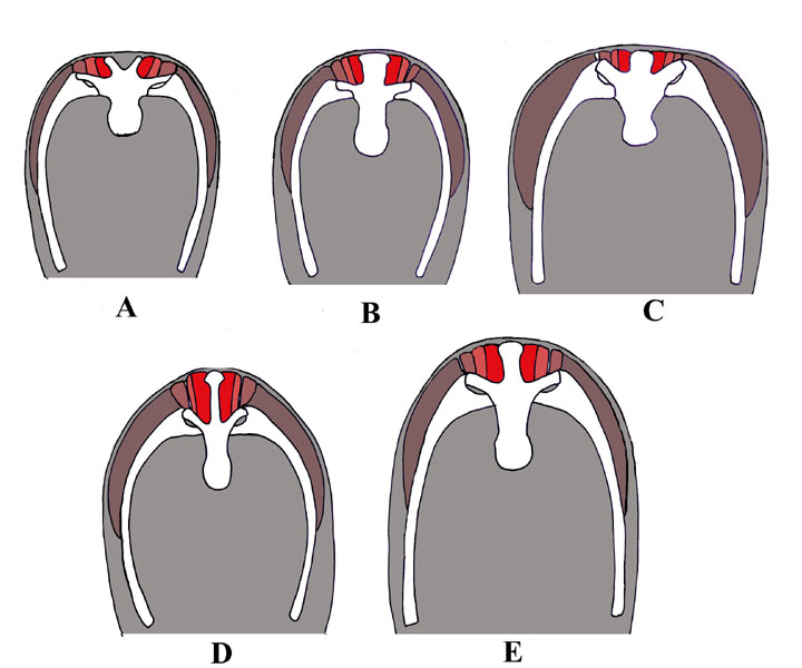
Figure 3) Cross section of the body of sauropods at the mid dorsal region; A-B) Camarasaurus; C) Saltasaurus; D) Shunosaurus; E) Haplocanthosaurus. Same muscle color identification as in figure 2.
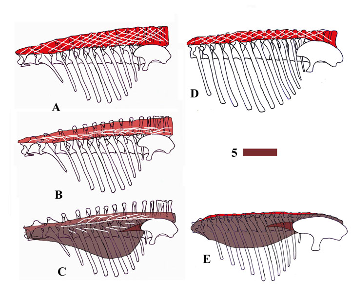
Figure 4) Side view of sauropods showing the muscles and tendons (white stripes); A-C Diplodocus; D) Camarasaurus; E) Saltasaurus. Same muscle color identification as in figure 2 with one more muscle; 5) ap ilcost.
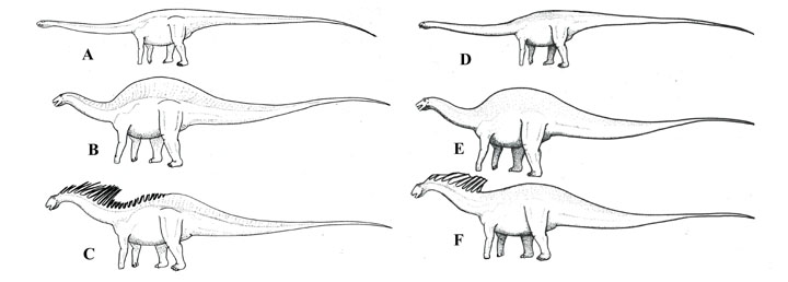
Figure 5) Side view restoration of sauropods, old way (A-C), new way (D-F);
A, D); B, E) Dicraeosaurus; C, F) Amargasaurus.
72) Ford, T. L., 2010, How to Draw Dinosaurs: Dull or colorful? What did dinosaurs look like? Prehistoric Times, n. 93, p. 18-19.
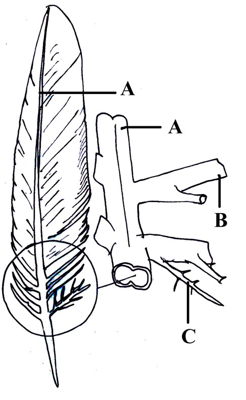
Figure 1) Feather diagram showing the placement of barbules. A) Shaft; B)
Barb; C) Barbules.
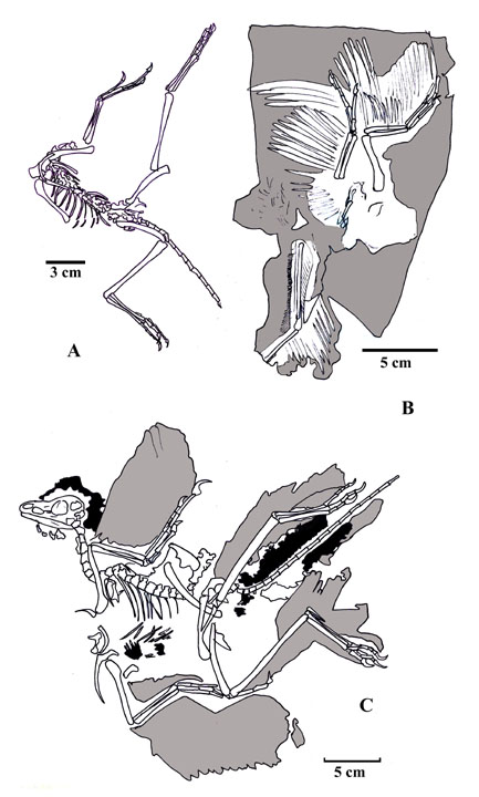
Figure 2) Skeletons of Anchiornis huxleyi; A) Type, IVPP V14378; B) Referred specimen BMHNC PH828; C) Referred specimen LPM-B00169.
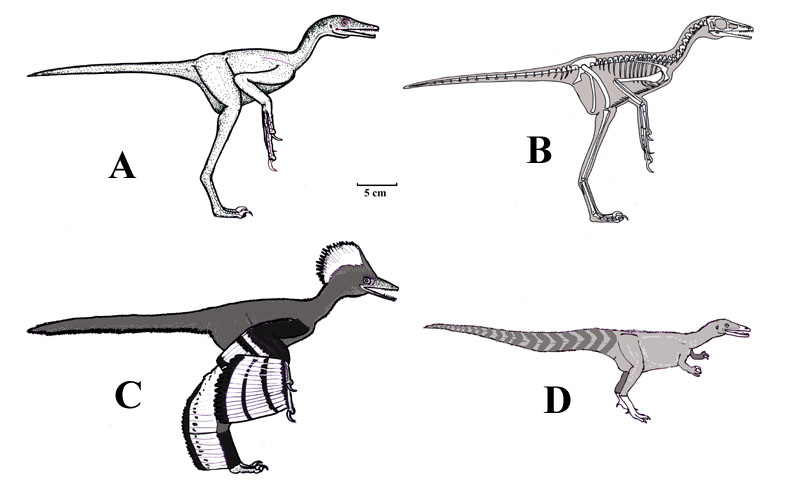
Figure 3) Reconstructions of Anchiornis and Sinosauropteryx;
A) Life restoration of Anchiornis (LPM-B00169) in the old version of scaly
dinosaurs; B) Skeleton
of referred specimen LPM-B00169; C) Life restoration of Anchiornis (LPM-B00169)
using recent studies showing the color pattern based on BMHNC PH828; D) Sinosauropteryx showing the color pattern on the tail.
72) Ford, T. L., 2010, How to Draw Dinosaurs: Aquatic Psittacosaurs (finally) Part one: Prehistoric Times, n. 94, p. 18-19.
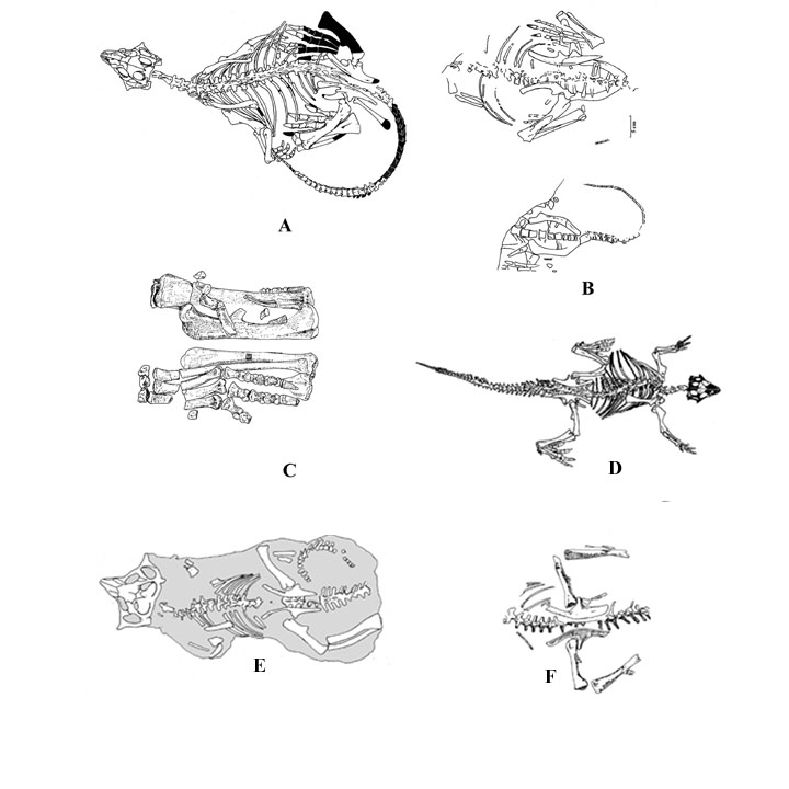
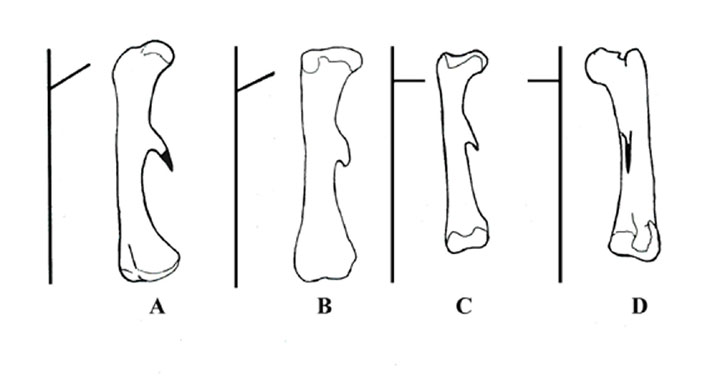
Figure 2) Ornithopod femurs. A) Psittacosaurus xinjiangensis:
B) Psittacosaurus
sibiricus; C) Hypsilophodon foxii; D) Iguanodon bernissartensis.
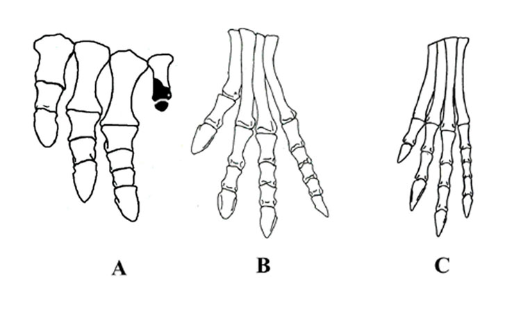
Figure 3) Manus and pes of psittacosaurs. A) Manus of Psittacosaurus sibiricus; B) Pes of Psittacosaurus neimongoliensis; B) Pes of Stenopelix valdensis.
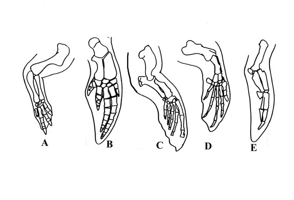
Figure 4) Front legs of swimming animals. A) Psittacosaurus; B) Dolphin (Lagenorhynchus);
C) Sea Lion (Zalophus); D) Sea Turtle (Cheloni); E) Penguin, (Spheniscus).
73) Ford, T. L., 2010, How to Draw Dinosaurs: Aquatic Psittacosaurs Part Two: Prehistoric Times, n. 95, p. 18-19.
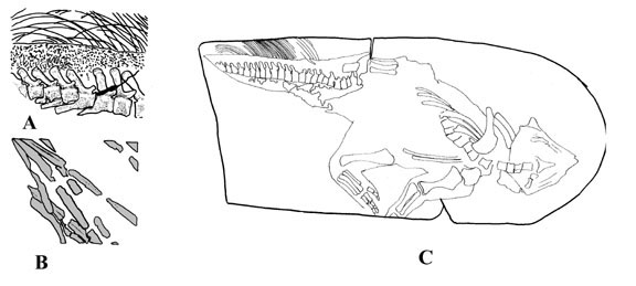
Figure 1), Bristal tail of Psittacosaurus.
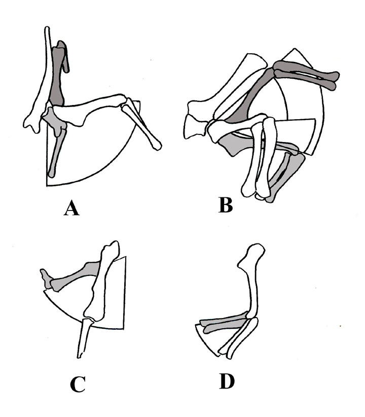
Figure 2). Range of movement of the pectoral girdle of Psittacosaurus neimongoliensis. A) cranial view; B) lateral view; C) dorsal view; D) lateral view of forelimb. All after Senter 2007.
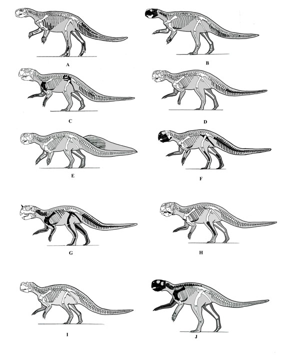
Figure 3). Psittacosaur skeletons in lateral view all to the same size. A) Psittacosaurus mongoliensis; B) Psittacosaurus mongoliensis = Protiguanodon mongoliensis; C) Psittacosaurus sinensis; D) Psittacosaurus neimongoliensis; E) Psittacosaurus sp (SMF R 4970); F) Psittacosaurus xinjiangensis; G) Psittacosaurus sinensis = Psittacosaurus youngi; H) Psittacosaurus major); I) Psittacosaurus sibiricus; J) Stenopelix valdensis.
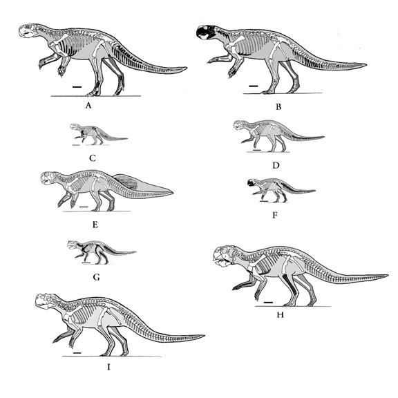
Figure 3). Psittacosaur skeletons in lateral view all to the same scale. A) Psittacosaurus mongoliensis; B) Psittacosaurus mongoliensis = Protiguanodon mongoliensis; C) Psittacosaurus sinensis; D) Psittacosaurus neimongoliensis; E) Psittacosaurus sp (SMF R 4970); F) Psittacosaurus xinjiangensis; G) Psittacosaurus sinensis = Psittacosaurus youngi; H) Psittacosaurus major); I) Psittacosaurus sibiricus.
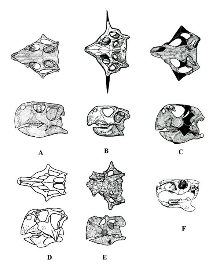
Figure 4). Psittacosaurus & capybara skulls. (A) Dorsal and lateral view
of Psittacosaurus mongoliensis (AMNH 6254) modified from Sereno et al. (1988).
(B) Psittacosaurus sinensis (IVPP V738), modified from Young (1958). (C) Psittacosaurus
meileyingensis (IVPP V7705), modified from Sereno et al. (1988). (D) Psittacosaurus
major (LH PV1), after Sereno et al. 2007. (E) Psittacosaurus sibiricus (PM
TGU 16/4-20), after Averianov et al. (2006). (F) Skull of capybara (free ware
illustration).
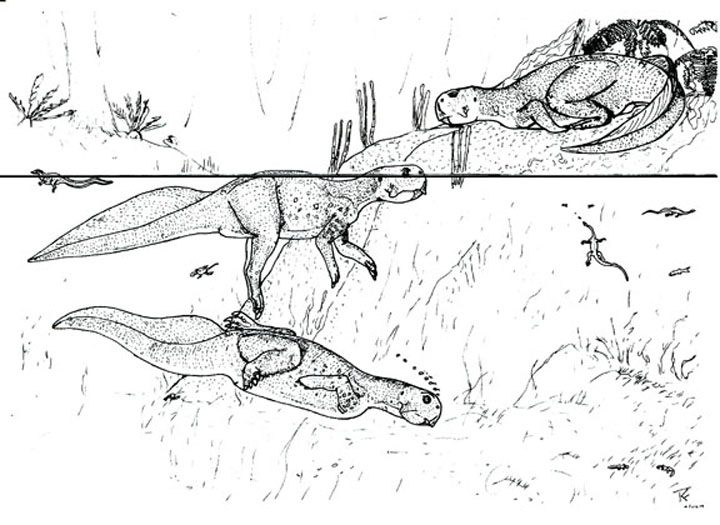
Figure 5). Life reconstruction of aquatic psittacosaurs (This wasn’t
used in the original article).
74) Ford, T. L., 2011, How to Draw Dinosaurs: To know the nose, Part 3 (Sauropods): Prehistoric Times, n. 96, p. 18-19.
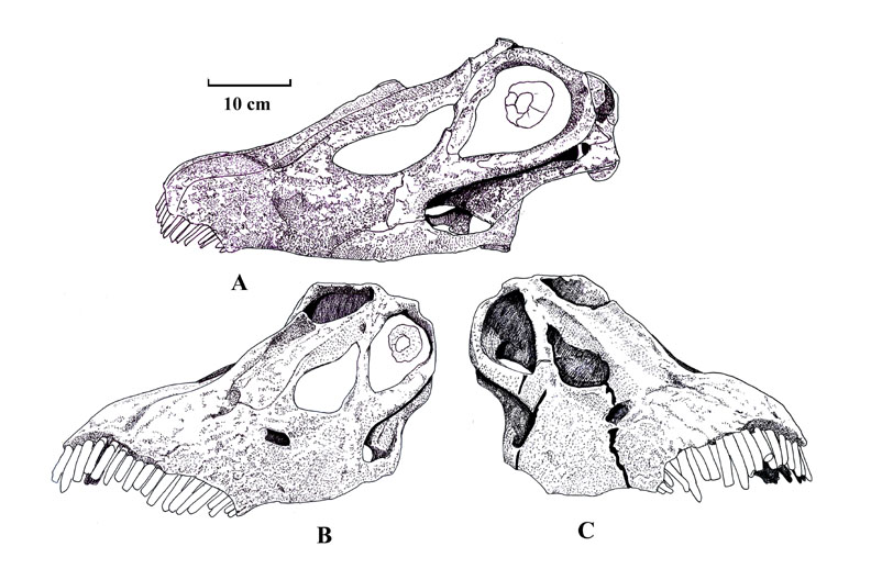
Figure 1) Skull of Diplodocus longus CM 11161; A) Side view; B) Left view; C) Right view.
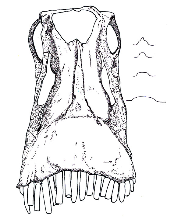
Figure 2) Skull of Diplodocus longus CM 11161. Front view and a schematic view of the ridge and shelf.
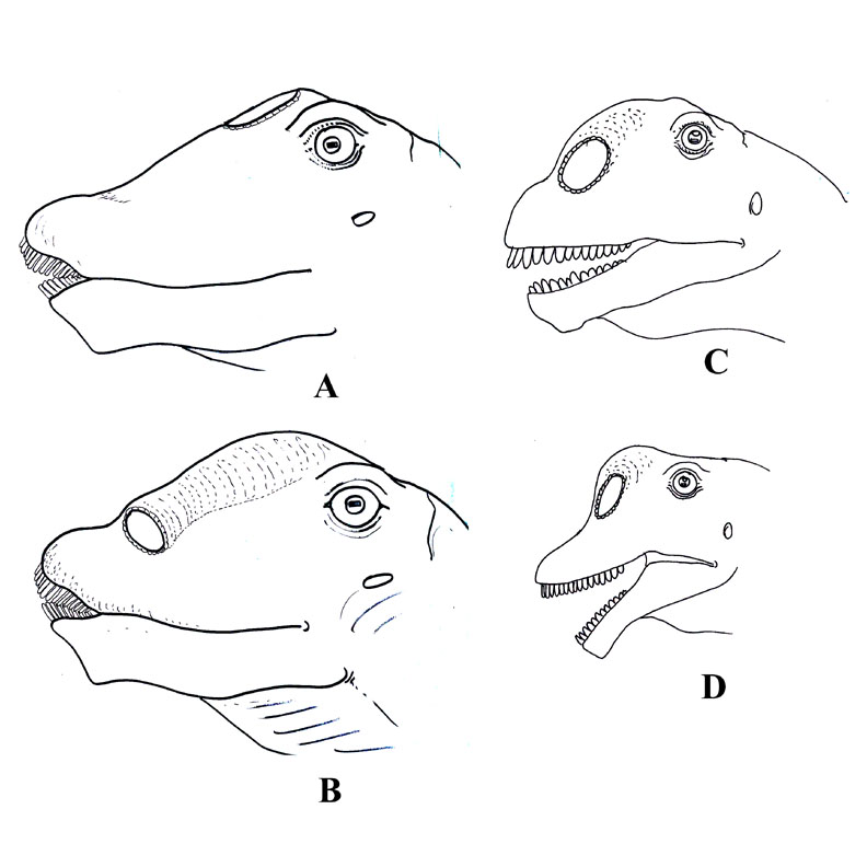
Figure 3) Head of Sauropods; A, B) Diplodocus longus CM 11161; A) Old interpretation of head and nose; B) New interpretation of nose and head; C) Camarasaurus head showing new interpretation of nose and head; D) Brachiosaurus sp from North America showing new interpretation of nose and head.
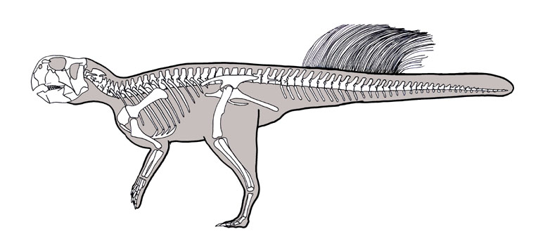
Figure 4) New interpretation of the skeleton and tail of Psittacosaurus based
on the cast of the Psittacosaurus with the bristle tail at the Carniege.
75) Ford, T. L., 2011, How to Draw Dinosaurs: And Now, The End of Dinosaurs: Prehistoric Times, no. 97, p. 18-19.
In figures 1-4 I’ll be using the same 6 dinosaurs. A) Deinonychus; B) Allosaurus; C) Tyrannosaurus; D) Hypsilophodon; E) Iguanodon; F) Gryposaurus. I modified Greg Pauls illustrations because he had both side and dorsal views of the skeletons.
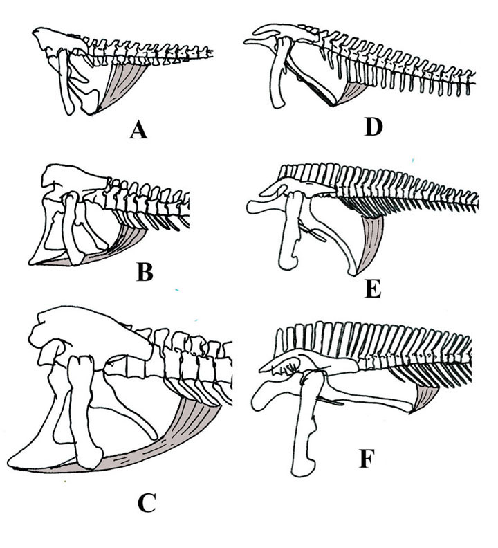 Figure 1) Caudotruncus muscle in Grey.
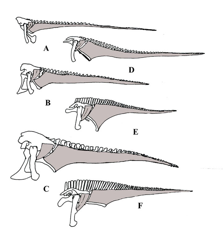
Figure 2) M. caudoformalis brevis which inserts on the 4th trochaner and attaches to the posterior ventral side of the ilium, and the M. ilio-ischocaudalis.
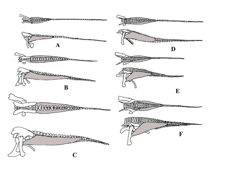
Figure 3) The m. caudofemoralis longus.
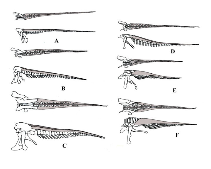
Figure 4) The M. longissimus and M. spinalis.
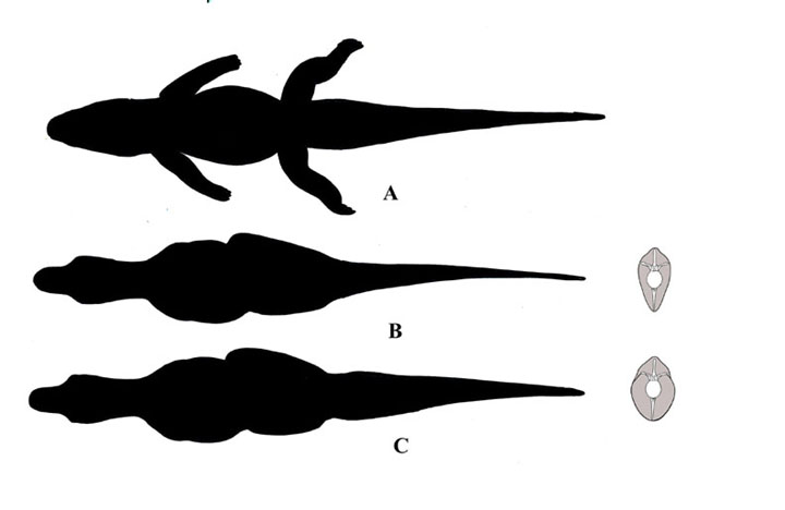
Figure 5) Silhouette illustrations of an Alligator A) and Tyrannosaurus
rex B) C). B) Showing the old version of a ‘thin’ tail with crossection
of Allosaurus tail modified from Madsen, 1976; C) New interpretation of a thicker
tail from Persons & Currie and a modified tail from Madsen, 1976.
76) Ford, T. L., 2011, How to Draw Dinosaurs: Bipedal or tripodal Sauropods? Prehistoric Times, v. 98, p. 18-20.
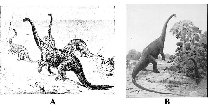
Figure 1): A) Amphicoelais altus restoration by Mr. Charles R. Knight under the direction of Prof. Cope, 1897; B) Diplodocus by Mr. Charles R. Knight , 1907.
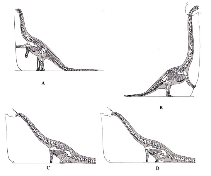 Figure 2): Tripodal sauropods; A) Diplodocus standing in a tripodal stance:
B) Omeisaurus tianfuensis (modified from Tanimoto, 1991); C) Kneeling sauropod
(modified from Siegwarth et al, 2010); D) Sitting sauropod (modified from Siegwarth
et al, 2010).
Figure 2): Tripodal sauropods; A) Diplodocus standing in a tripodal stance:
B) Omeisaurus tianfuensis (modified from Tanimoto, 1991); C) Kneeling sauropod
(modified from Siegwarth et al, 2010); D) Sitting sauropod (modified from Siegwarth
et al, 2010).
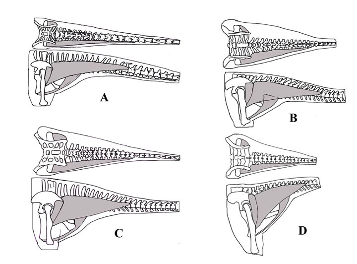 Figure 3): Tail muscles in sauropods, m. caudoformalis brevis which inserts
on the 4th trochanter and attaches to the posterior ventral side of the ilium,
the m. caudoformalis longus, and the caudotruncus which inserts on the pubis,
slides along the ischum and attaches to caudal chevrons: A) Diplodocus; B)
Camarasaurus: C) Apatosaurus: D) Brachiosaurus (all
modified after Paul).
Figure 3): Tail muscles in sauropods, m. caudoformalis brevis which inserts
on the 4th trochanter and attaches to the posterior ventral side of the ilium,
the m. caudoformalis longus, and the caudotruncus which inserts on the pubis,
slides along the ischum and attaches to caudal chevrons: A) Diplodocus; B)
Camarasaurus: C) Apatosaurus: D) Brachiosaurus (all
modified after Paul).
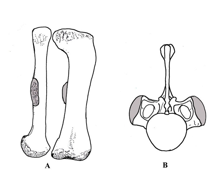 Figure 4): Femur and caudal vertebrae showing muscle attachment area (in grey);
A) Femur of Brontosaurus excelsus showing 4th trochanter; B) Brontosaurus
excelsus second caudal vertebra in anterior view showing muscle attachment area.
Figure 4): Femur and caudal vertebrae showing muscle attachment area (in grey);
A) Femur of Brontosaurus excelsus showing 4th trochanter; B) Brontosaurus
excelsus second caudal vertebra in anterior view showing muscle attachment area.
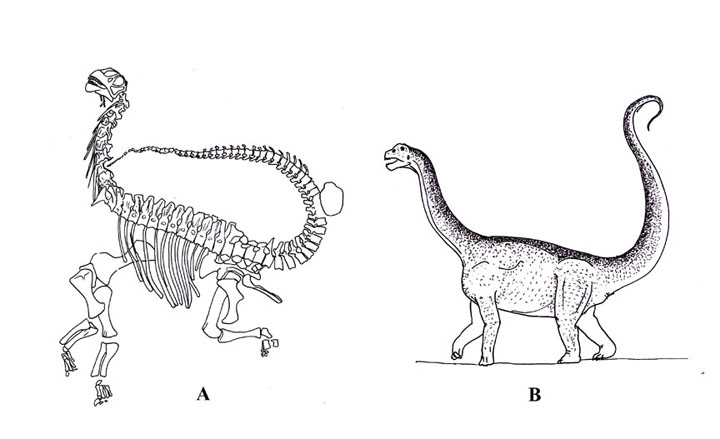 Figure 5): Camarasaurus; A) Camarasaurus lentus juvenile specimen (CM 11338)
from Dinosaur National Monument. Modifed from Gilmore 1925); B) Camarasaurus with a vertical tail (just to see what it could look like).
Figure 5): Camarasaurus; A) Camarasaurus lentus juvenile specimen (CM 11338)
from Dinosaur National Monument. Modifed from Gilmore 1925); B) Camarasaurus with a vertical tail (just to see what it could look like).
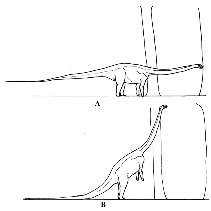 Figure 6): Diplodocus standing with a tree in front of it and one at its shoulder.
A) standing quadrapedally; B) bipedal/tripodal Diplodocus showing how far it
would have to walk to the tree in front of it so it could brace itself on the
trunk, and to show that it would have to twist its body to the closer tree.
Figure 6): Diplodocus standing with a tree in front of it and one at its shoulder.
A) standing quadrapedally; B) bipedal/tripodal Diplodocus showing how far it
would have to walk to the tree in front of it so it could brace itself on the
trunk, and to show that it would have to twist its body to the closer tree.
77) Ford, T. L., 2011, How to Draw Dinosaurs: Terror Birds, not so terrible...: Prehistoric Times, no. 99, p. 18-19.
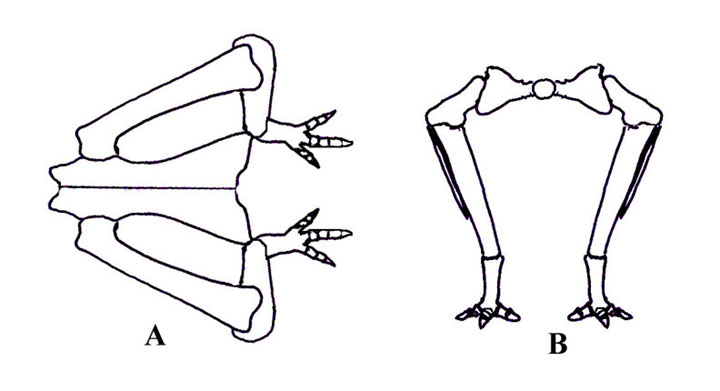
Figure 1) A schematic illustration of how a birds femur. A) Dorsal view showing the femora bowing away from the body; 2). Cross-section of the pelvic area showing how the femora is bowed.
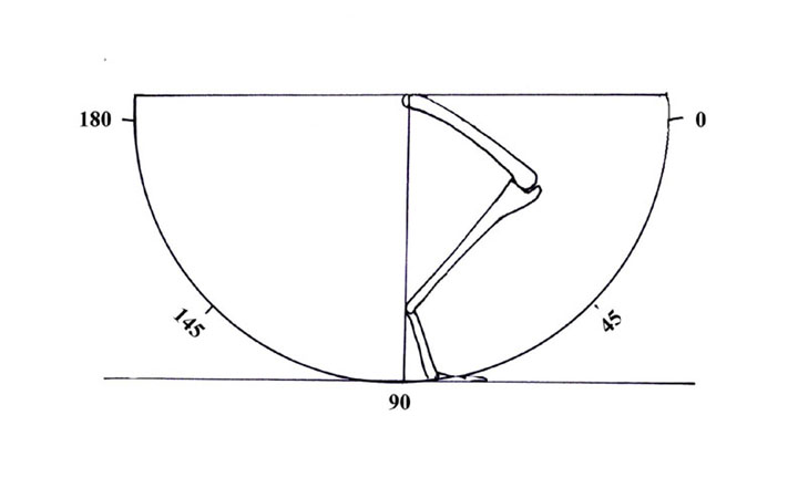
Figure 2) Schematic of the arch used in the locomotion of birds and dinosaurs
used in the paper.
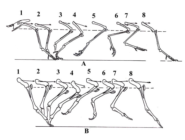
Figure 3) Stages of locomotion in birds. A) Gatsey’s Guinea fowl locomotion; B) Murray & Vickers-Rich Mihirungs locomotion. The striped line is the ‘knee’ line, the arrows point the direction of the femur is going.
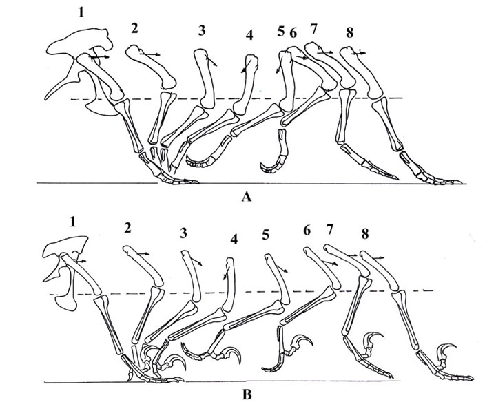
Figure 4) Stages of locomotion in theropods. A) Tyrannosaurus rex; B) Deinonychus.. The striped line is the ‘knee’ line, the arrows point the direction of the femur is going.
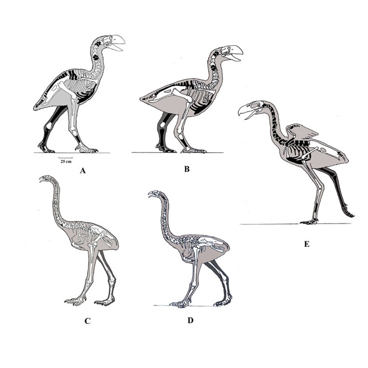
Figure 5) A) Diatryma typical illustration; B) New interpretation of Diatryma;
C) Moa typical illustration; D) New interpretation of a Moa; E) New interpretation
of Patagornis marshi.
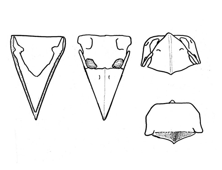
Figure 6). New interpretation of the skull of Diatryma.